Dilip Kumar Tiwari*, Kajal Sharma, Shivani Goyal, Kaushelendra Mishra, Abhishek Chaturvedi
Lakshmi Narain College of Pharmacy, Bhopal 462022, Madhya Pradesh, India
*Address for Corresponding Author
Dilip Kumar Tiwari
Lakshmi Narain College of Pharmacy, Bhopal
Raisen Road Bhopal-462022, Madhya Pradesh, India
Abstract
Background: Diabetes mellitus (DM) is one of the leading metabolic disorders worldwide. Diabetes mellitus is due to a lack of function of β pancreatic cells or insensitivity of target tissue to insulin. This disorder not only affects the metabolic system but also causes various associated problems like neuropathy, nephropathy, retinopathy, and heart-related disease. Treatment of Diabetes mellitus (DM) with allopathic drugs for a long duration has been not safer and may cause several adverse effects along with hypoglycemia. World health organization (WHO) recommended drug researchers emphasize herbal-based research which is safer and more effective for longer regimens in dosing. Objective: This work was aimed to attempt to explore roots extract of Aegle marmolus for antidaibetic potential by in vitro methods. Material and methods: In this research work Aegle marmolus roots extract were tested for anti-diabetic potential by in vitro method α–amylase inhibitory assay and Glucose uptake in yeast cells. Results and conclusion: Results were shown α-amylase inhibitory activities IC50 112 μg/ml. EEAMR exhibited highest percentage of glucose uptake i.e. 54.84±0.752, which was almost near to the standard i.e. 61.74±0.527. In conclusion antidiabetic effect of Aegle marmolus is attributed to the mixture of phytochemicals in the extracts. In present study extract showing excellent antidiabetic potential by in-vitro method. Possible activities due to presence of bioflavonoid & flavonoid constituents present in extract which mimic the insulin activity & glucose uptake by adipose tissue.
Keywords: diabetes mellitus, hypoglycemia, neuropathy, retinopathy, α-amylase, Aegle marmolus
Introduction
Diabetes mellitus is recognized as being a syndrome, a collection of disorders that have hyperglycemias and glucose intolerance as their hallmark, due either to insulin deficiency or to the impaired effectiveness of insulin’s action, or to a combination of these (Albright, 1997). In order to understand diabetes it is necessary to understand the normal physiological process occurring during and after a meal. Food passes through the digestive system, where nutrients, including proteins, fat and carbohydrates are absorbed into the bloodstream (Stewart, 1999).
The human pancreas is basically composed of two types of secretory cells that are both involved in nutrient handling: 98% of the cells- the exocrine type – secrete a food- processing enzyme-bicarbonate mixture into the duodenum, while the remaining 2% - the endocrine type. The islet changes, from a morphological point of view, associated with various types of diabetes can be divided into those with and without severe beta-cell loss. Severe beta-cell loss is found in type I diabetes and some uncommon forms of diabetes such as virus-related diabetes and congenital diabetes. Islets without severe loss of beta-cells are encountered in type II diabetes and in the secondary forms of diabetes (Kloppel et al., 1997).
Unfortunately, there is no cure for diabetes yet but by controlling blood sugar levels through a healthy diet, exercise and medication the risk of long-term diabetes complications can be decreased. Medicinal plants which have showed anti-diabetic activity during earlier investigations include Panax species, Phyllanthus species, Acacia arabica, Aloe vera, Aloe barbadensis, Artemisia pallens, Momordica charantia, Alium cepa, Trigonella foenum-graecum etc.
Materials and methods
Aegle marmelos root obtained from LNCP garden by digging the root & then it was thoroughly washed and dried under shade. Plant prior authenticated by Suman Mishra scientist Vindhya herbal Bhopal. The further drug is cut into small pieces coarsely powdered and pulverized. The coarse powder is then extracted by petroleum ether and then ethyl acetate by the maceration process.Further, it was phytochemically investigated for the presence of various phytoconstituents category.
Preparation of Ethanolic extract of Aegle marmelos:
The powdered plant materials were extracted with 70% v/v ethanol by hot continuous percolation method using soxhlet apparatus (Harborne, 1984). The solvent from the extract was removed under reduced pressure using rotary evaporator and subjected to freeze drying in a lyophilizer until dry powder was obtained. The extract of Aegle marmelos was stored in air tight container and utilized.
Qualitative phytochemical analysis (Van et al., 2011; Soni et al., 2011)
The extracts obtained by successive solvent extraction were subjected to various qualitative tests to detect the presence of chemical components
Evaluation of antidiabetic activity by using in vitro assays
Alpha-amylase inhibitory assay (Brenyah, 2013)
The Alfa–amylase inhibitory assay of ethanolic extact of Aegle marmolus root was evaluated according to a previously described method by (Ranilla et al., 2008) . In brief, 0.5 ml of extract was mixed with 0.5 ml of α-amylase solution (0.5 mg/ml) with 0.02 M sodium phosphate buffer (pH 6.9 with 0.006 M NaCl). The mixture was incubated at room temperature for 10 min and 0.5 ml of starch solution (1%) in 0.02 M sodium phosphate buffer (pH 6.9 with 0.006 M NaCl) was added. The resulting mixture was incubated at room temperature for 10 min, and the reaction was terminated using 1 ml of dinitrosalicylic acid color reagent. At this time, the test tubes were placed in a water bath (100 °C for 5 min) and cooled until room temperature was attained. The mixture was then diluted with 10 ml of deionized water, and absorbance was determined at 540 nm. The absorbance of blank (buffer instead of extract and amylase solution) and control (buffer instead of extract) samples were also determined. Acarbose was used as standard drug.
The inhibition of α-amylase was calculated using the following equation

Where Abs control corresponds to the absorbance of the solution without extract (buffer instead of extract) and with α -amylase solution
Abs sample corresponds to the solution with extract and α -amylase solution
Glucose uptake in yeast cells (Madeleine, 2022)
Glucose uptake assay by yeast cells was performed according to (Cirillo et al., 1963) . The yeast, (Saccharomyces cerevisiae) suspended in distilled water was subjected to repeated centrifugation (3000 × g, 5 min) until clear supernatant fluids were obtained and 10% (v/v) of the suspension was prepared in distilled water. Various concentrations of plant extracts (50 to 250 μg/ml) were added to 1 ml of glucose solution (5 mM) and incubated together for 10 min at 37 °C. Reaction was started by adding 100 μl of yeast suspension followed by vortexing and further incubation at 37 °C for 60 min. After 60 min, the tubes were centrifuged (2500 × g, 5 min) and amount of glucose was estimated in the supernatant. Acarbose was used as standard drug. The percentage increase in glucose uptake by yeast cells was calculated using the following formula:

Where, Abs sample is the absorbance of test sample and Abs control is the absorbance of control reaction (containing all reagents except the test sample). All the experiments were carried out in triplicates.
Capsule Formulation (King et al., 1993)
EEAMR was taken for the formulation development. The crude extract of Aegle marmolus was blended with the various excipients for convenient to formulate as a unit dosage form as capsules and then evaluated. It was carried out with EEAMR-300mg/capsule and cross povidone XL10 as super disintegrant used in the formulation, which is presented in Table 4. The bioactive compounds and excipients were passed through 60 # sieve, weighed and mixed well. Finally, the preservatives were mixed and filled in “0” size hard gelatin capsule using hand filling machine with the average weight of 450 mg per capsule.
Evaluation of the capsule formulation (Chadwick et al., 2007)
Uniformity of Weight
An intact capsule was weighed separately and the capsule was opened without any loss or part of the shell and removed the contents as completely as possible. The weight of the content was calculated from the difference between the capsule with content and the shell weight alone.
Moisture Content
Moisture content was determined by using automatic Karl Fischer titration apparatus.
Disintegration Test
The test was carried out on 6 capsules using tablet disintegration tester ED-20 (Electrolab, Mumbai, India) distilled water at 37°C±2°C was used as a disintegration media and the time in second taken for complete disintegration of the capsule with no palable mass remaining in the apparatus was measured in sec.
Microbiological Test (Davis and Granner, 1996)
The formulated capsules were subjected to microbiological evaluation, which was carried out as per the procedures of Indian pharmacopoeia, 2007 and WHO Guideline.
I. Total viable count
II. Test for absence of Escherichia coli
III. Test for absence of Salmonella sp.
Bacterial Count
To about 9 – 10cm diameter petridish containing 15ml of liquefied soya bean casein digest agar maintained at not more than 45°C, One ml of pre-treated sample was added. The petridish were incubated at 37°C for 48h and the numbers of colonies were counted (Mutel et al., 2011).
Test for absence of E. coli
One ml of pre-treated sample was added to the tube containing 5ml of Mac Conkey Broth. The tube was incubated at 42°C for 48h, and observed for development of pink color (Esmaeili, 2021).
Test for Absence of Salmonella sp.
Each 10g of sample was taken in 2 different conical flasks. In the first conical flask 100ml of pre sterilized peptone buffer was added. In the second conical flask 100ml of fluid lactose broth was added. From each conical flask 5ml of sample was withdrawn and 100ml of pre-sterilized nutrient broth was added. The nutrient broths with samples were incubated at 35°C for 24h. At this end of the time, one ml of the nutrient broth was added to tetra thionate brilliant green bile broth and the tubes were incubated at 35°C for 48h. After that bile broth was transferred into bismuth sulphate agar plate. In another tube, one ml of bile broth was transferred into brilliant green agar plate. Both the plates were incubated at 35°C for 48h. The colour of bismuth sulphate agar plate changed from green to black metallic color colony. It indicates positive and the brilliant green agar plate turns green to pink color colony (Gerich, 2001).
Analysis of drug content
Quantification of Kaempherol-3-O-Glucoside equivalent to Kaempherol by HPTLC The analysis is performed with HPTLC (Camag, Switzerland). The active principle is applied with the linomat IV applicator on the HPTLC silica gel 60 F 254 plates (E-Merck, Germany). The plates are developed with a twin-trough developing chamber. After 70 developments, the plates are scanned with a Camag TLC scanner 3 and the data are processed with Win CATS software. The HPTLC analysis of the active principle is carried out using different solvent systems for individual plant components (WHO, 1985)
Result and discussion
Table 1. Phytochemical tests for EEAMR
|
TESTS |
Observations |
|
Alkaloids |
|
|
Mayer’s test |
+ ve |
|
Wagner test |
+ ve |
|
Hager’s test |
+ ve |
|
Carbohydrates And |
|
|
Molisch test |
+ ve |
|
Legal’s test |
+ ve |
|
Borntrager’s test for anthraquinones |
- ve |
|
Glycosides |
|
|
Legal test |
- ve |
|
Keller Kiliani Test |
- ve |
|
Terpenoids & Phytosterols |
|
|
Liebermann-Burchard test |
+ ve |
|
Salkowski test |
+ ve |
|
Flavonoids |
|
|
Shinoda test |
+ ve |
|
Fluorescence test |
+ ve |
|
Tannins & Phenolic compound |
|
|
Ferric chloride test |
+ ve |
|
Potassium dichromate test |
+ ve |
|
Lead acetate test |
+ ve |
|
Proteins |
|
|
Millon’s test |
- ve |
|
Biuret Test |
- ve |
|
Ninhydrin Test |
- ve |
|
Saponins |
|
|
Saponification test |
- ve |
|
Fats & Lipids |
|
|
Spot test |
- ve |
Alpha-amylase inhibitory assay
The ethanolic extact of Aegle marmolus root were subjected to α-amylase inhibitory assay along with Acarbose as a standard drug. The ethanolic extract showed higher activity among all other extracts tested (Fig-1) which was comparable to standard acarbose. The α-amylase inhibitory activities of differed solvent extracts are recorded in Table 2.
Table 2. α -Amylase inhibitory activities and IC50 values by Aegle marmolus root extracts
|
Sample |
Concentration |
Inhibition I % |
IC50(μg/ml) |
|
Ethanol extract |
100 μg/ml |
48.264± 0.457 |
112 μg/ml |
|
Standard (Acarbose) |
100 μg/ml |
56.752± 0.312 |
86.22 μg/ml |

Figure 1. Percentage inhibition of α-amylase by Aegle marmolus root extracts.
Glucose uptake in yeast cells
Ethanolic extract of Aegle marmolus root extracts are subjected to in vitro glucose uptake assay employing yeast as model. The percentage of glucose uptake in yeast cells by the extract was compared with standard drug Acarbose. Aqueous extract exhibited higher activity than the remaining solvent extracts tested (Fig .2). There was concentration dependent increase in percentage of glucose uptake with increasing in concentration of Aegle marmolus extract (5.4). Ethanolic extract exhibited highest percentage of glucose uptake i.e. 54.84±0.752, which was almost near to the standard i.e. 61.74±0.527 (Fig.2) at 250 μg concentration. Results also indicated that Aegle marmolus had almost same efficiency in increasing the glucose uptake by yeast cells as compared to standard drug Acarbose.
Table 3. Percentage of glucose uptake in yeast cells treated with Aegle marmolus ethanolic
root extracts
|
Samples |
Concentration(μg/ml) |
Inhibition (%) |
IC50(μg/ml) |
|
Standard |
50 μg |
42.23±0.241 |
48.65 μg |
|
100 μg |
46.52±0.513 |
||
|
150 μg |
49.31±0.362 |
||
|
200 μg |
55.25±0.421 |
||
|
250 μg |
61.74±0.527 |
||
|
Ethanol extracts |
50 μg |
35.12±0.378 |
154.87 μg |
|
100 μg |
37.25±0.642 |
||
|
150 μg |
43.74±0.258 |
||
|
200 μg |
48.67±0.325 |
||
|
250 μg |
54.84±0.752 |

Figure 2. Percentage of glucose uptake in yeast cells treated with Aegle mormolus extracts.
Table 4. Working Formulas of EEAMR capsules
|
S. No |
Ingredients |
Weight (mg) |
|
|
EEAMR |
300 |
|
|
Dicalcium phosphate anhydrous |
105 |
|
|
Sodium lauryl sulphate |
10 |
|
|
Cross povidone XL 10 |
10 |
|
|
Light magnesium stearate |
20 |
|
|
Methyl paraben |
4 |
|
|
Propyl paraben |
1 |
Table 5. Physiochemical parameters of capsule
|
S. No |
Name of the test |
Observation |
|
1. |
pH |
7.52 |
|
2. |
Moisture Content |
3.62%w/w |
|
3. |
Weight Variation (Mean ± SEM) |
472± 5 mg |
|
4. |
Disintegration Time (Mean ± SEM) |
5 Min 45 Sec± 0.32 |
Table 6. Microbiological evaluation of capsule
|
S. No |
Name of The Test |
Limit of Herbal Formulation as Per WHO |
Observation |
|
|
Total microbial count bacterial count (Per gram of sample) |
Aerobic bacteria max105/g |
874 CFU/g |
|
|
Fungal count( per gram of sample ) |
Moulds maximum 103/g |
360 CFU/g |
|
|
Presence of escheria colli |
Should be absent |
Absent |
|
|
Presence of salmonella Sp |
Should be absent |
Absent |
Quantification of Rutin derivative in EEAMR by HPTLC
Quantification of Rutin derivative in EEAMR by HPTLC assay as described in Schematic representation of the HPTLC chromatograms of Rutin derivative equivalent to Rutin EEAMR are observed in Figure 3 and Figure 4 and under UV 254nm and UV 366nm the HPTLC plates, which are shown in Figure 5. Rf value and AUC values are shown in Table 6.

Figure 3. HPTLC peaks of standard Rutin and EEAMR in 3D view
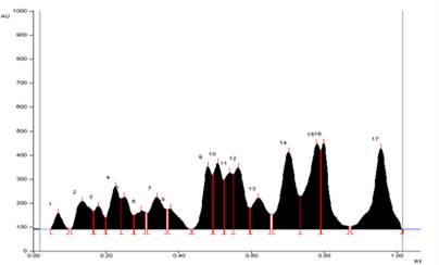
Figure 4. HPTLC chromatogram of standard Rutin and EEAMR
Conclusion
The antidiabetic effect of Aegle marmolus is attributed to the mixture of phytochemicals in the extracts. The phytochemicals responsible for antidiabetic properties mainly are alkaloids, phenolic acids, flavonoids, glycosides, saponins, polysaccharides, stilbenes, and tannins. In the several animal studies reported using different plants, there is a wide variety between the extractions methods. In present study extract of showing excellent antidiabetic potential by in-vitro method. Possible activities due to presence of bioflavonoid & flavonoid constituents present in extract which mimic the insulin activity & glucose uptake by adipose tissue.
Conflict of interest
Author declare that no conflict of interest
References
Albright AL. 1997. Diabetes In: Exercise Management for persons with chronic diseases and disabilities. USA: Human Kinetics (Braun-Brumfield). pp 94-100.
Stewart DL, Stewart DL. 1999. The Baltimore Alliance for the Prevention and Control of Hypertension and Diabetes: a model for developing a colorectal cancer community outreach program. Journal of the Association for Academic Minority Physicians. 10(3):77-9
van Huyssteen M, Milne PJ, Campbell EE, van de Venter M. 2011. Antidiabetic and Cytotoxicity Screening of Five Medicinal Plants Used by Traditional African Health Practitioners in the Nelson Mandela Metropole, South Africa. African journal of traditional, complementary, and alternative medicines: African Networks on Ethno medicines. 8. 150-8.
Deutschländer, MS, Deutschländer, MS, Namrita Lall, Maryna van de Venter. 2010. Isolation and identification of a novel anti-diabetic compound from Euclea undulata Thunb. South African Journal of Botany, 76:393-394.
Brenyah RC, Ephraim RKD, Owiredu WKBA, Eghan BA, Quaye JnrL. 2013. Prevalence and determinants of proteinuria among type 2 diabetics in Kumasi, Ghana Journal of Medical and Biomedical Sciences, 2(1):13-21
“Madeleine Albright” 2022. Britannica, The Editors of Encyclopaedia. . Encyclopedia Britannica, 11 May.
Yajnik CS. 1992. Diabetes secondary to tropical calcific pancreatitis, Baillière's clinical endocrinology and metabolism, volume 6(4) pages 777-796
King H, Rewers M. 1993. Global estimates for prevalence of diabetes mellitus and impaired glucose tolerance. WHO ad hoc diabetes reporting group. diabetes care, 16:157-177.
Gerich JE, Gerich JE. 2010. Diabetic Medicine. Feb; 27(2):136-42
Mutel E, Gautier-Stein A, Abdul-Wahed A, Amigó-Correig M, Zitoun C, Stefanutti A, Houberdon I, Tourette JA, Mithieux G, Rajas F, Mutel E. 2011. Diabetes Dec; 60(12):3121-31
Chadwick WA. Roux S, Van de Venter, M LouwJ, Oelofsen W. 2007. Anti- diabetic effects of Sutherlandia frutescens in Winstar rats fed a diabetogenic diet. Journal of Ethnopharmacology, 109. pp121 -127
Davis SN, Granner DK. 1996. Insulin, oral hypoglycaemic agents and the pharmacology of the endocrine pancreas in Goodman and Gilman’s The Pharmacological basis of Therapeutics 9th edition. Hardman J.G. and Limbird L.E. The McGraw-Hill Companies Inc, USA. pp. 1487-1518.
Gerich JE. 2001. Matching Treatment to Pathophysiology in type 2 Diabetes. Clinical Therapeutics. Vol.23, No 5. pp. 646-659
Grover JK, Yadav S, Vats V. 2002. Medicinal plants of India with anti-diabetic potential. Journal of Ethnopharmacology. 81(1): 81-100
World Health Organization study group on diabetes mellitus. 1985. Technical report series No. 727. World Health Organization, Geneva.pp.1-108
Soni H, Ribadiya C, Bhatt B, Sheth N. 2010. Evaluation of herbal formulation (capsule) containing Ashwagandha as a single herb with their nutritional value determination. International Journal of Applied Biology and Pharmaceutical Technology, 1:960-967.
Esmaeili S, Dayani L, Taheri A, Zolfaghari B. 2021. Phytochemical standardization, formulation and evaluation of oral hard gelatin capsules from Pinus eldarica bark extract. Avicenna Journal of Phytomedicine. Mar-Apr; 11(2):168-179
Klöppel G, In’T Veld PA. 1997. Morphology of the Pancreas in normal and diabetes states. In: International Textbook of Diabetes Mellitus, Second Edition. K.G.M.M Alberti, P. Zimmet, R.A. DeFronzo and H. Keen. John Wiley and Sons Ltd. New York.pp. 287-313.


